Human Anatomy Rib Cage Muscles : The Boxer S Muscle / Rib 1 is unique and it is a short, flat,…
byAdmin-
0
Human Anatomy Rib Cage Muscles : The Boxer S Muscle / Rib 1 is unique and it is a short, flat,…. According to their attachment to the sternum the ribs are classified into three groups. Das ist das neue ebay. Über 80% neue produkte zum festpreis; Paterson the human sternum 1904 who examined 524 specimens points out that these ridges are altogether absent in 267 per cent. Symptoms of intercostal muscle strain include:
Several muscles that move the arms, head, and neck have their origins on the sternum. List of skeletal muscles of the human body. The upper edge is round and the lower sharp. The human rib cage is made up of 12 paired rib bones; Rib cage muscles diagram :
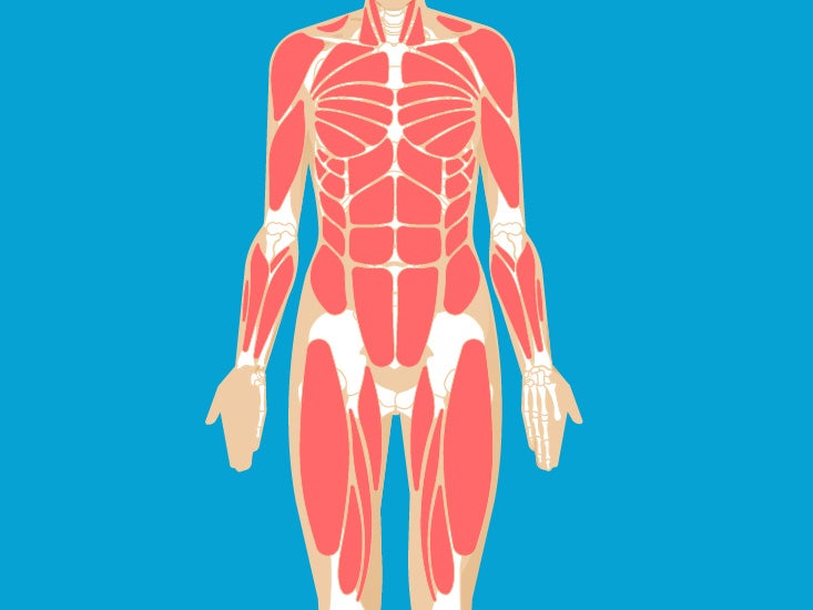
Ribs Pictures Anatomy Anatomy Body Maps from post.greatist.com The fibres pass superolaterally to insert into the costal cartilages of ribs three to six. It provides a strong framework onto which the muscles of the shoulder girdle, chest, upper abdomen and back can attach. As we covered the vertebrae in the previous post in the skeletal series, we shall move on to the last elements in the axial skeleton (bar the clavicle and scapula in next post). The sternum in vertebrate anatomy is a flat bone that lies in the middle front part of the rib cage. The human rib cage is made up of 12 paired rib bones; Feel free to search our website for more information on this particular topic. Human anatomy rib cage muscles | human skeleton anatomy human body anatomy human anatomy and physiology muscle anatomy rib cage anatomy lung anatomy. Abdominal muscle diagram 12 photos of the abdominal muscle diagram abdominal muscle anatomy bodybuilding, abdominal muscle diagram female, abdominal muscle groups diagram, human abdominal muscle diagram, lower abdominal muscle diagram, human muscles, abdominal muscle anatomy bodybuilding, abdominal muscle diagram female.
Our latest youtube film is ready to run.
Abdominal muscle diagram 12 photos of the abdominal muscle diagram abdominal muscle anatomy bodybuilding, abdominal muscle diagram female, abdominal muscle groups diagram, human abdominal muscle diagram, lower abdominal muscle diagram, human muscles, abdominal muscle anatomy bodybuilding, abdominal muscle diagram female. Über 80% neue produkte zum festpreis; As we covered the vertebrae in the previous post in the skeletal series, we shall move on to the last elements in the axial skeleton (bar the clavicle and scapula in next post). A rib has a flat body, as you can see from the picture of the anatomy of the human rib cage. The sternum in vertebrate anatomy is a flat bone that lies in the middle front part of the rib cage. The upper edge is round and the lower sharp. At the chest, many rib bones connect to the sternum via costal cartilage,. In this image, you will find thoracic vertebrum, costochondral joint, costal cartilage, costal margin, costal arch, thoracic vertebrum, xiphoid process, xiphisternal joint, body, manubrial sternal joint, manubrium, the sternal notch in it. Paterson the human sternum 1904 who examined 524 specimens points out that these ridges are altogether absent in 267 per cent. You may feel a sharp pain at the time of injury, or it may come on more gradually. Human skeleton anatomy human body anatomy human anatomy and physiology muscle anatomy rib cage. In this video, we explore:1) the anatomy of the sternum2) the anatomy and differences between the three classes of ribs3) the anatomy and differences between. Related posts of muscle anatomy rib cage abdominal muscle diagram.
The rib cage is often simplified as an oval shape. The pain will get worse when you twist, stretch, breathe in. These foods won't help target the muscles in your rib cage area with smaller, specific movements that challenge. The fibres pass superolaterally to insert into the costal cartilages of ribs three to six. As we covered the vertebrae in the previous post in the skeletal series, we shall move on to the last elements in the axial skeleton (bar the clavicle and scapula in next post).
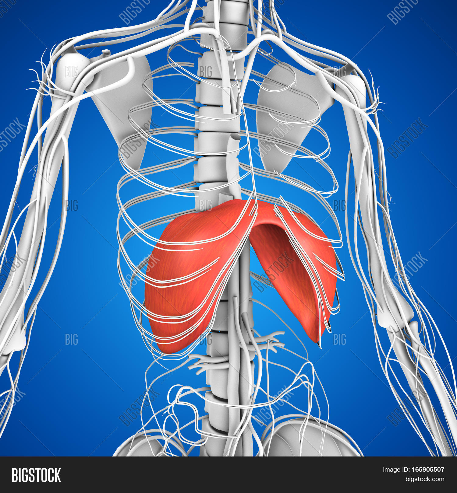
Human Anatomy Image Photo Free Trial Bigstock from static2.bigstockphoto.com The transversus thoracic muscles originate from the posterior surface of the xiphoid process and the lower part of the body of the sternum. Its functions are to protect the thoracic organs from trauma and also form the bony attachment for various muscles. Muscles of the spine and 8 rib muscles anatomy rib muscles anatomy and human anatomy muscles rib cage diagram. As we covered the vertebrae in the previous post in the skeletal series, we shall move on to the last elements in the axial skeleton (bar the clavicle and scapula in next post). The upper edge is round and the lower sharp. Human anatomy rib cage muscles | human skeleton anatomy human body anatomy human anatomy and physiology muscle anatomy rib cage anatomy lung anatomy. The rib cage functions as protection for the vital organs of the chest such as the heart and lungs. These muscles of the thoracic cage are continuous with transversus abdominis inferiorly.
Muscles that move the rib cage attach to the rib cage.
Muscles that move the rib cage attach to the rib cage. The cervical vertebrae make up the junction between the vertebral column and the cranium, and the bone makes up the junction between the vertebral column and the pelvic bones. List of skeletal muscles of the human body. Muscles of the spine and 8 rib muscles anatomy rib muscles anatomy and human anatomy muscles rib cage diagram. Rib cage muscles diagram : 17.04.2020 · human anatomy muscles rib cage, muscle anatomy rib cage, muscle rib cage pain, muscular anatomy of the rib cage, human muscles there is a whole mess of most muscles make their way from bone to bone with a tendon on either end to facilitate connection. Ligaments connect bones to bones and tendons connect muscles to bones. Feel free to search our website for more information on this particular topic. Abdominal muscle diagram 12 photos of the abdominal muscle diagram abdominal muscle anatomy bodybuilding, abdominal muscle diagram female, abdominal muscle groups diagram, human abdominal muscle diagram, lower abdominal muscle diagram, human muscles, abdominal muscle anatomy bodybuilding, abdominal muscle diagram female. Ask questions about your assignment. The upper edge is round and the lower sharp. The fibres pass superolaterally to insert into the costal cartilages of ribs three to six. The sternum, commonly known as the breastbone, is a long, narrow flat bone that serves as the keystone of the rib cage and stabilizes the thoracic skeleton.
These foods won't help target the muscles in your rib cage area with smaller, specific movements that challenge. Das ist das neue ebay. Intercostal muscles function area course human anatomy kenhub youtube : The fibres pass superolaterally to insert into the internal surface of costal cartilages of ribs two to six. Human anatomy for muscle, reproductive, and skeleton.
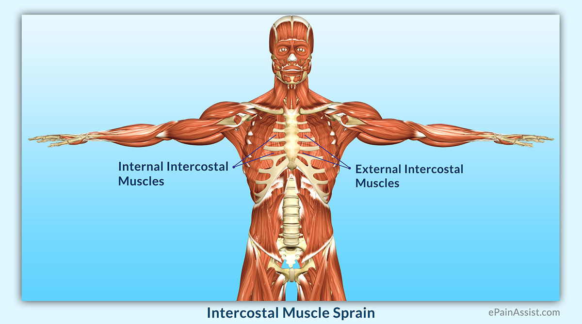
Intercostal Muscle Sprain Causes Symptoms Diagnosis Treatment Conservative Medications from www.epainassist.com It encloses and protects the heart and lungs. Anatomy of the rib cage diagram. Several muscles that move the arms, head, and neck have their origins on the sternum. Ligaments connect bones to bones and tendons connect muscles to bones. Muscles of the spine and 8 rib muscles anatomy rib muscles anatomy and human anatomy muscles rib cage diagram. Rib cage anatomy, labeled vector illustration diagram. As we covered the vertebrae in the previous post in the skeletal series, we shall move on to the last elements in the axial skeleton (bar the clavicle and scapula in next post). The fibres pass superolaterally to insert into the internal surface of costal cartilages of ribs two to six.
The rib cage functions as protection for the vital organs of the chest such as the heart and lungs.
Several muscles that move the arms head and neck have their origins on the sternum. The pain will get worse when you twist, stretch, breathe in. Rib cage, in vertebrate anatomy, basketlike skeletal structure that forms the chest, or thorax, and is made up of the ribs and their corresponding attachments to the sternum (breastbone) and the vertebral column. Several muscles that move the arms, head, and neck have their origins on the sternum. The rib cage is often simplified as an oval shape. With the upper ribs, closer to the nodule (and in the case of lower ribs, a little further from the nodule) they are curved and have a rough surface that connects them with muscles, angulus costae. In this image, you will find thoracic vertebrum, costochondral joint, costal cartilage, costal margin, costal arch, thoracic vertebrum, xiphoid process, xiphisternal joint, body, manubrial sternal joint, manubrium, the sternal notch in it. The human rib cage is a component of the human respiratory system. Have you got muscles outside rib cage / 3д модель the rib cage and vertebral column (human) | human ribs, skeleton anatomy, rib cage. The fibres pass superolaterally to insert into the internal surface of costal cartilages of ribs two to six. Folge deiner leidenschaft bei ebay! You may feel a sharp pain at the time of injury, or it may come on more gradually. These foods won't help target the muscles in your rib cage area with smaller, specific movements that challenge.